Blood Vessels Labeled Brain : (a) and (b) the main blood vessels supplying the brain in ... - Equal to the intestinal muscles that move the food morsel along brain level:
Blood Vessels Labeled Brain : (a) and (b) the main blood vessels supplying the brain in ... - Equal to the intestinal muscles that move the food morsel along brain level:. Only some of the vessels that exist in a real brain have been labeled. If you use this material, please attach a link to the artwork, i would really love to see it :d. Blood vessels are intricate networks of hollow tubes that transport blood throughout the entire body so that it can deliver valuable nutrients to and remove waste from cells. The difference in the structural characteristics of arteries, capillaries and veins is attributable to their respective functions. Label the blood vessels of the male pelvis using the hints provided.
The precise relation between blood vessels and brain regions, reflecting the physiology and pathology of brain function directly and accurately, has figure 3. Another whole article within the blood vessels and csf section is dedicated to the cavernous sinus. These vessels transport blood cells, nutrients, and oxygen to the tissues of the body. The brain and its surrounding blood vessels exist in a close relationship. About 2 years ago updated:
Blood vessel - Wikipedia from upload.wikimedia.org They also take waste and carbon dioxide away from the tissues. Label the blood vessels in the inferior view of the brain using the hints provided. The blood vessels are the components of the circulatory system that transport blood throughout the human body. The blood vessels (and nerves) enter the brain through holes in the skull called foramina. He says the restricted vessels prevent the blood from draining fast enough and injure the brain by causing a build up of iron which leads to ms. This is particularly important structure due to its clinical implications, which are discussed in more detail in the article. The brain and its surrounding blood vessels exist in a close relationship. The brain and its surrounding blood vessels exist in a close relationship.
Over 1 year ago version:
The blood vessels (and nerves) enter the brain through holes in the skull called foramina. Equal to the intestinal muscles that move the food morsel along brain level: Ultrasound may offer a safe way to more as the name suggests, this is a barrier between the brain's blood vessels (capillaries) and the cells and other components that make up brain tissue. This vessel supplies blood to the front part of your brain, knows as your frontal lobe. • identification of blood vessels as arteries, capillaries or veins from the structure of their walls. Label the blood vessels of the male pelvis using the hints provided. The difference in the structural characteristics of arteries, capillaries and veins is attributable to their respective functions. Red indicates arteries, and blue. Over 1 year ago version: About 2 years ago updated: Blood vessels are vital for the body and play a key role in diabetes helping to transport glucose and insulin. Blood vessels innervate all tissues in vertebrates, enabling their survival by providing the necessary nutrients, oxygen, and hormonal signals. The two cell types ensure the integrity of the neural vasculature by maintaining the blood.
This is particularly important structure due to its clinical implications, which are discussed in more detail in the article. • identification of blood vessels as arteries, capillaries or veins from the structure of their walls. Blood travels from the heart in arteries, which branch into smaller and smaller vessels, eventually becoming arterioles. The structure, distribution and labeling of the whole brain vascular system of different arteries and veins in 3d. Posterior communicating a internal carotid а.
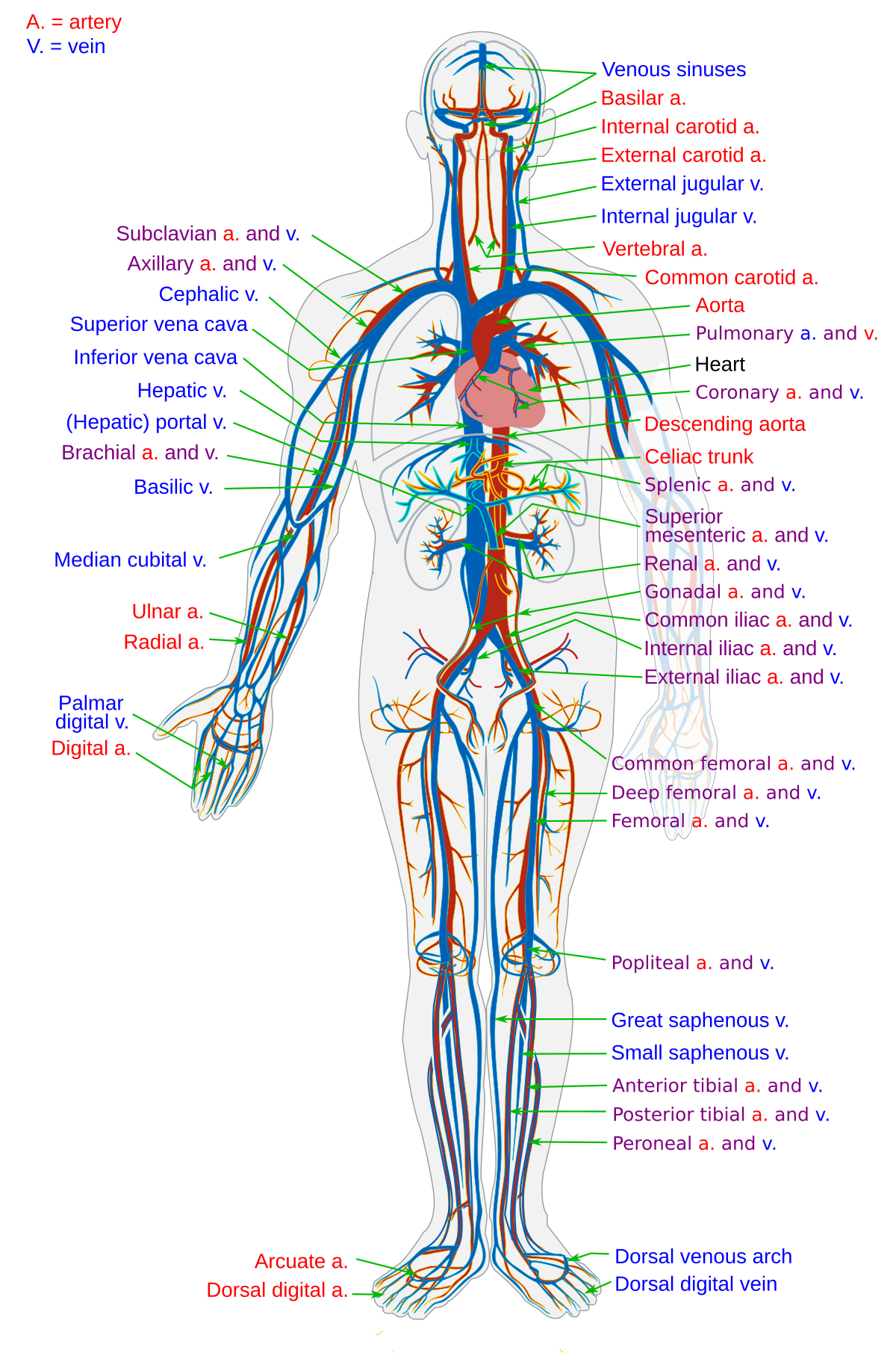
blood vessels Flashcards | Easy Notecards from www.easynotecards.com The 500 ms patients, both adults and children, also underwent mri scans of the brain to measure iron deposits in surrounding areas of the brain. Equal to the intestinal muscles that move the food morsel along brain level: This vessel supplies blood to the front part of your brain, knows as your frontal lobe. This is particularly important structure due to its clinical implications, which are discussed in more detail in the article. Blood is also supplied to the brain by the vertebral a. Blood vessels 2 labeled palmar arch digital artery right femoral a right femoral v great saphenous vein left popliteal a right anterior tibial a. Fill in the blanks with the appropriate words to describe blood flow from the heart. Label the blood vessels in the inferior view of the brain using the hints provided.
In the cerebral medulla, the arteries and veins of the right side of the body are controlled from the left side of the brain;
Blood vessels are intricate networks of hollow tubes that transport blood throughout the entire body so that it can deliver valuable nutrients to and remove waste from cells. The difference in the structural characteristics of arteries, capillaries and veins is attributable to their respective functions. Blood vessels are referred to collectively as the vascular system and, together with the heart, make up the circulatory system or cardiovascular system. Label the blood vessels in the inferior view of the brain using the hints provided. He says the restricted vessels prevent the blood from draining fast enough and injure the brain by causing a build up of iron which leads to ms. Equal to the intestinal muscles that move the food morsel along brain level: Blood travels from the heart in arteries, which branch into smaller and smaller vessels, eventually becoming arterioles. Learn vocabulary, terms and more with flashcards, games and other study tools. Supplies the anterior brain and the vertebral a. Only some of the vessels that exist in a real brain have been labeled. As well as providing new insights into the. Supplies the posterior brain, blood supply to the entire brain is ensured by anastomoses between the vessels. The dense tight junctions between endothelial cells prevent paracellular transport through the.
Label the blood vessels in the inferior view of the brain using the hints provided. Equal to the intestinal muscles that move the food morsel along brain level: The blood vessels are the components of the circulatory system that transport blood throughout the human body. The precise relation between blood vessels and brain regions, reflecting the physiology and pathology of brain function directly and accurately, has figure 3. Researchers have discovered how cells of the blood vessels sense the metabolic condition of the brain and alter vascular function in response.
16. Blood vessels/flow - Anatomy & Physiology Biol121 with ... from classconnection.s3.amazonaws.com Growing brains in laboratories was just the start for scientists. The structure, distribution and labeling of the whole brain vascular system of different arteries and veins in 3d. In the cerebral medulla, the arteries and veins of the right side of the body are controlled from the left side of the brain; In the article on the ventricles within the cns, we will discuss their structure and. The 500 ms patients, both adults and children, also underwent mri scans of the brain to measure iron deposits in surrounding areas of the brain. The two cell types ensure the integrity of the neural vasculature by maintaining the blood. He says the restricted vessels prevent the blood from draining fast enough and injure the brain by causing a build up of iron which leads to ms. Internal carotid artery (anterior circulation), vertebral artery (posterior circulation), and their hexagonal anastomotic network called blood brain barrier refers to the wall between the brain tissue and blood vessels.
In the cerebral medulla, the arteries and veins of the right side of the body are controlled from the left side of the brain;
Label the blood vessels in the inferior view of the brain using the hints provided. The 500 ms patients, both adults and children, also underwent mri scans of the brain to measure iron deposits in surrounding areas of the brain. Microscopically, it is formed by the endothelium of the blood vessel. Blood vessels 2 labeled palmar arch digital artery right femoral a right femoral v great saphenous vein left popliteal a right anterior tibial a. Blood vessels are vital for the body and play a key role in diabetes helping to transport glucose and insulin. As well as providing new insights into the. Red indicates arteries, and blue. Comes off the subclavian a., ascends although the internal carotid a. The blood vessel wall is endowed with connective tissue, smooth muscle, and striated muscles. The precise relation between blood vessels and brain regions, reflecting the physiology and pathology of brain function directly and accurately, has figure 3. Learn vocabulary, terms and more with flashcards, games and other study tools. Over 1 year ago version: These vessels transport blood cells, nutrients, and oxygen to the tissues of the body.
Blood vessels innervate all tissues in vertebrates, enabling their survival by providing the necessary nutrients, oxygen, and hormonal signals blood vessels labeled. Supplies the posterior brain, blood supply to the entire brain is ensured by anastomoses between the vessels.


0 Komentar